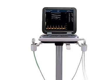
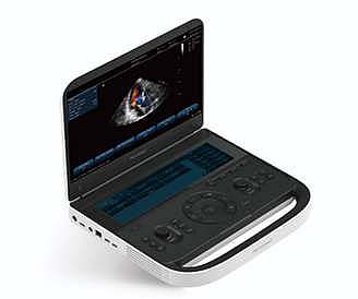
Advanced Imaging Boosts Diagnostic Confidence
- B, B+B, 4B, B+M, M
- Pulse doppler (PW)
- Anatomical 3M-mode (AM)
- B+PW (real-time dual synchronization)
- Tissue harmonic imaging (THI),
- Pulsed inverse harmonic imaging (ITHI)
- Tissue specific imaging (TSI)
- Puncture enhancement (optional)
- Wide-field imaging (optional)
- Contrast imaging (optional)
- 3D/4D imaging (optional)
Optimized Parameters Enhance Diagnostic Accuracy
- 15″ high definition LCD, tilt display
- Quick Save One push image transfer to local or directly to USB
- Stored images can be edited and measured again
- Picture-in-picture, partial zoom function
- POpti™ Auto Optimization: single button image optimization
- PStation™: clipboard, onboard reporting
- IMT: auto-measurement of carotid intimae-thickness
TSI
TSI’s tissue-specific imaging function sets dedicated sound velocity matching for different examination parts, such as muscles, liquids, fats, etc., to improve the diagnostic value of obese patients or special diagnosis parts, making the ultrasound image layer clearer and the particles finer., the resolution is better.
PureSight Imaging Technology
Our PureSight™ technology reduces speckle artifacts, improves smoothness along preferred edge directions, and maintains consistent local average gray levels. This results in clearer and more visually appealing images, enhancing diagnostic confidence.
PrismX™ Spatial Composite Imaging
By employing PrismX™ spatial composite imaging technology, our system enhances contrast, fine resolution, and spatial resolution. It optimizes tissue and pathological interface echo continuity while minimizing artifacts such as specular reflection, speckle, scattering, and attenuation, ensuring superior image clarity.
AMM
Anatomic M-type echocardiography can also be used to diagnose complex heart diseases, such as congenital heart disease, valvular heart disease, etc. Through detailed observation and analysis of the heart structure, doctors can more accurately judge the type and extent of the disease and develop a more appropriate treatment plan for patients.
PanoramaVision™ Extended Field Imaging
PanoramaVision™ technology captures a series of two-dimensional sectional images, which are reconstructed into an ultra-wide sectional image with a continuous field of view. This enables precise measurement of organ size and lesions, while providing a comprehensive view of the lesion’s scope, intemal echo, location, size, and adjacent tissues.


 Convex+ transvirginal + cardiac probe
Reviews
There are no reviews yet.