


Feature:
Comprehensive and professional dry eye examination and diagnosis program
Support 7 ways of dry eye detection functions to provide reliable basis for clinical diagnosis.
The test report with pictures and texts , the test results shown clearly and in detail.
A full set of dry eye examinations can be completed in 7 minutes, effectively improving clinic efficiency.
Non-invasive inspection method, safer operation, patient comfort and easy cooperation.
Fully automated software analysis + Quantification of testing standards, easier diagnosis by doctors.
Convenient patient management system, which can continuously record the development of patients.
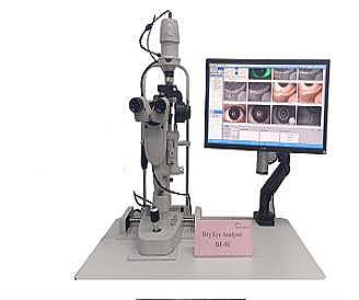
Dry eye project inspection report
Rich in content, 7 items of dry eye examination data are fully displayed on one report.
Be clear at a glance, Sufficient image and data combination, clear and detailed display of inspection results.
Multiple templates, Support single report, any combination of multiple projects.
TMH
Comfortable and reliable.
Using infrared light with a wavelength of 850μm as the light source,
it will not stimulate the secretion of tears,
the patient is more comfortable and the result is more reliable.
Automatic identification, identification, measurement.
Ocular redness analysis
Conjunctiva, sclera, two modes.
Multi-dimensional analysis of the patient’s ocular surface hyperemia.
Provide more evidence for diagnosis.
Automatic identification, analysis, and classification.
Gland Opening
HD eyelid image. Can clearly obtain the overall shape of the eyelid margin.
Image with slight changes in gland openings.
Corneal Staining
Professional yellow filter.
Make corneal fluorescein sodium stained image clearer.
Automatic identification, analysis, classification
NIBUT
Comfortable and reliable: The use of infrared rays with a wavelength
of 850μm will not stimulate tear secretion, and record the maintenance
time of the tear film in the original state of the eye, and the result is
more reliable.
All-round: Provides first and average break-up time, tear film break-up
diagram, and graph to display the situation of tear film break in all round.
Lipid layer analysis
The white ring projection can completely present the true color
and shape of the lipid layer.
Automatic identification, analysis, and classification
Using professional AI algorithm, lipid layer thickness
measurement can be accurate to 10nm
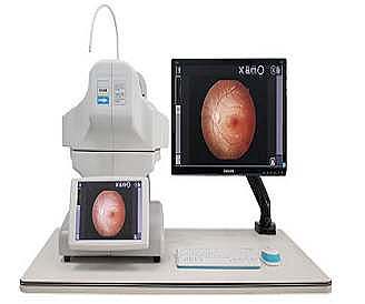
Meibomian glands imaging analysis
Clear. AI image enhancement.
Professional infrared imaging can clearly observe the
morphology of the meibomian acini.
Support automatic identification, strengthen the contrast of glands, and automatically calculate the proportion of meibomian glands missing.

Reviews
There are no reviews yet.