

Feature:
. The collector has an high depth of field effect, which ensures
one image can capture more tissue lesions. Supplemented by
front diaphragm controller, enjoy more depth of field effects.
Using PWM pulse modulation to control the brightness of the
light source regardless of the brightness, always ensure the
stability of the light source color.
Rich picture post processing function meet the clinical
requirements
Intuitive support for one click export. data transfer is more
convenient.
Soft coaxial background light satisfies the operator before
photography
Based with step less dimming, which can fine adjust the
brightness,
more comfortable for the client; can control the brightness
with one hand, more convenient operation.
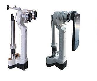
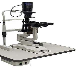
Infrared/ultraviolet filters are specially designed in the slit
lamp lighting, which can protect the eyes of the doctor and
patient in daily use.
Technical Data
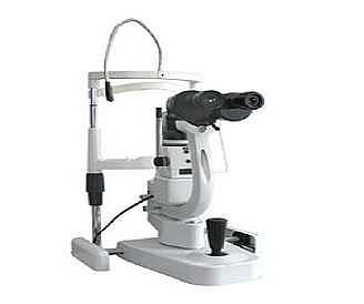
Optical design type: Galileo type
Magnification Changer: Revolving Drum
Eyepiece: 12.5X
Magnification: 6X, 10X, 16X, 25X, 40X
Diopter compensation range: +7D
Interpupillary distance range: 52mm-85mm
Visual diameter: 6x(ρ33): 10x(p22); 16x(φ14) ;25x(φ8.5) : 40x(q5.5)mm
Slit width: 0mm-14mm
Slit height: 1mm~14mm
Slit angle horizontal rotation: 0°~180° Slit Tilting Angle 5°, 10°, 15°, 20°
Spot diameter: φ0.2, φ1, φ3, φ5, φ10, φ14(mm)
Color filters: heat absorption, grey filter, red-free, cobalt blue
Light source: Warm color LED bulb
Light control mode: Step less dimming on the base
Digital indication mode: cross type dividing panel
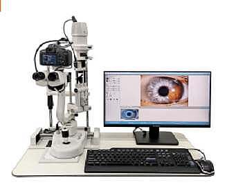
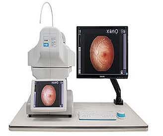
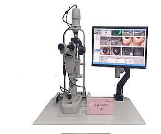
Digital collector: built-in professional level medical collector with 20 million pixels
Background light: Coaxial background light, embedded adaptive software
Image acquisition, Automatic recognition of OD&OS, Image processing
Software system: Image marking, Image comparison, Print the report, Export the image


Reviews
There are no reviews yet.